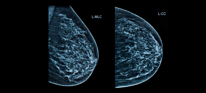
Digital Mammography

ARYA STANA BONE & BREAST CARE has Cape Town’s and the Western Cape’s most up to date Digital Mammography technology including CAD (Computer Aided Detection) and 3D Breast Tomosynthesis with the new Clarity HD – High Resolution Imaging Technology & The SmartCurve Stabilization System to transform the breast imaging patient experience.
The SmartCurve system provides a curved compression surface that offers a more comfortable patient experience without compromising image quality, exam time, dose or workflow. It improved comfort in 93% of patients who reported moderate to severe discomfort with standard compression.
Digital mammography (special x ray of breast tissue) offers higher resolution and higher quality images compared to traditional mammograms taken on film (analog mammogram).
Digital mammography is the most technologically advanced imaging tool available for the early detection of breast abnormalities in women with a history of Breast Cancer or a new breast symptom (diagnostic mammography) or for an annual routine check in women without breast symptoms (screening mammography).
Digital image capture improves contrast, shortens exam time (10-15 minutes) and allows image exchange with other Doctors without information loss. Images are both obtained and stored electronically eliminating lost films, thereby improving interval screening with availability of previous mammograms for comparison. Because the images can be adjusted by the breast radiologist, more subtle abnormalities may be noted.
Every woman who has a Digital Mammogram at ARYA STANA BONE & BREAST CARE is provided with a ‘second read’ using CAD. After the breast radiologist reads your mammogram, CAD technology is used as a ‘second read’ of the original mammogram, with suspicious areas highlighted for the breast radiologist to review again. CAD technology has been proven to increase the accuracy of mammography.
Breast Tomosynthesis is a method of acquiring and displaying three dimensional (3D) mammograms that provide greater image clarity than conventional two dimensional (2D) mammograms by reducing the confounding effects of superimposed breast tissue.
Breast Tomosynthesis is a 3D imaging technology that involves acquiring images of a stationary compressed breast at multiple angles during a short scan. Individual images are then reconstructed into a series of thin high resolution slices that can be displayed individually or in dynamic cine mode.
At ARYA STANA BONE & BREAST CARE we are dedicated to promoting women’s health through early detection and diagnosis of Breast Cancer. Early detection provides the best chance to treat Breast Cancer successfully. Routine annual mammography is the only screening method that has been proven to be effective in early detection of Breast Cancer.
An annual mammogram for Breast Cancer screening is recommended for all women from the age of 40 and earlier for those who have any concern or a family history of Breast Cancer.
ARYA STANA BONE & BREAST CARE with the latest technology in Digital mammography and experienced, caring staff will provide you the best care you deserve.
Frequently Asked Questions
What is digital mammography?
ARYA STANA BONE & BREAST CARE is the first and only site in the Western Cape’s private health sector to offer Digital Mammography with computer-aided detection (CAD) and 3D Tomosynthesis. Digital mammography is similar to standard mammography in that x-rays are used to produce detailed images of the breast. The difference is that digital mammography is equipped with a digital receptor and generates computerized images immediately instead of a film cassette that needs to be developed into a film. A similar comparison is a standard camera to a digital camera.
What is large field of view digital mammography?
At ARYA STANA BONE & BREAST CARE we want to make your mammogram as comfortable as possible. Our Selenia Dimensions full-field digital mammography system is recognized as the technology leader in the mammography market place and is being used in leading well women’s clinics around the world.
Similar to traditional digital mammography systems the large field of view is equipped with a larger paddle providing a more comfortable mammogram. The larger paddle allows for more precise imaging, especially for women with large breasts. The larger paddle results in fewer compressions, less radiation and less pain and fewer retakes.
What is computer-aided detection or CAD?
Computer aided detection or CAD is a sophisticated computer program that is linked to the digital mammography system and that has been shown in studies to increase the accuracy of mammography by up to 20%. After the radiologist has processed the digital breast images on the monitor and done the interpretation, CAD is activated. The system scans the images and alerts the radiologist to take a second look by flagging any potentially suspicious areas. The radiologist then reviews these areas again to determine if they need further study. CAD is like having a second set of trained eyes reviewing every mammogram. By detecting early or subtle changes, CAD can allow for earlier intervention and greater chances for cure.
What is Breast Tomosynthesis?
Breast Tomosynthesis is a revolutionary technology that gives radiologists the ability to identify and characterise individual breast structures without the confusion of overlapping tissue. During a Tomosynthesis scan, multiple, low-dose images of the breast are acquired at different angles. These images are then used to produce a series of one millimeter thick slices that can be viewed as a three dimensional reconstruction of the breast.
Instead of viewing all tissue complexities on a traditional 2D mammogram, the breast radiologist can now scroll through the layers of the breast in one-millimeter thick slices. This allows the breast radiologist to see around features in the tissue and identify areas of concern that may have been hidden by overlapping tissue, or dismiss normal areas that may have appeared suspicious on a digital mammogram. As a result, recalls may be reduced, unnecessary biopsies may be eliminated, and breast cancers may be identified earlier.
Image quality is key to early detection of Breast Cancer.
What is the difference between traditional (analog) and digital mammography?
Analog mammography uses x-ray to record images on film using an x-ray cassette. Films are then “developed” and produced and put on a light box and read by the Radiologist. With Digital mammography the x-rays produce a digital image on a screen while the patient is still in position. The technologist has the ability to review these in “real time” to determine image quality. Once completed the images are sent to the radiologist electronically at a reading station where they can manipulate, view and magnify areas of breast tissue. This enhances the information available for reading and interpretation. From the patient’s perspective there is little difference because the exam is conducted in a similar way except that the exam is shorter in length. Compression of the breast is required for both digital and analog mammography.
Will you be able to compare past analog mammograms to digital mammograms?
In either case, the images can be compared from exam to exam and from digital to analog.
Is digital a better technology?
While analog mammography is still a sound and reliable exam, digital mammography offers a new ability to process and view the images. This results in shorter exam time for the patient, and greater flexibility for the radiologist in interpreting the images. Much like a digital photo, the images can be enhanced, manipulated, and improved by the radiologist, so a digital mammogram can provide more information for diagnosis. Digital technology also offers better visibility of the entire breast. The capabilities of digital mammography result in fewer repeat views, which means less patient exposure.
What if I have dense breasts?
If you have dense breast tissue it is likely that digital mammography will provide better imaging quality for you with reduced radiation dose. This will be reviewed based upon your history and previous mammograms.
When will I learn the results of my digital mammogram?
Once your exam is complete, our dedicated breast radiologist will carefully evaluate the images and meticulously compare your current mammogram with your previous mammograms in looking for any slight change. At your appointment, the breast radiologist will ensure that all necessary examinations are performed and will discuss the findings with you before you leave. You will receive a written report at the end of your consultation and a written report will be sent to your Doctor.
If you require a follow up appointment this may be scheduled before you leave or at your convenience.
What can I expect?
During a digital mammogram your experienced certified mammographer (technologist) will normally obtain two X-rays images of each breast. Additional images may be necessary. Accuracy requires precise identical positioning of the breast in the mammography device and adequate compression of the breast tissue (more on this later). Position and compression are required to ensure that the images are of the highest quality and that all the breast tissue is imaged. The entire exam will take approximately 10-15 minutes.
Compression (Will my mammogram be painful?)
Compression of the breast tissue is vital to the accuracy of the exam. For most women, a mammogram is not painful, merely uncomfortable – and only for the seconds that their breasts are compressed. For comfort sake it is best to schedule your mammogram 7-10 days after the onset of your last menstrual cycle.
How to prepare on the day of your mammogram?
On the day of your mammogram, it is best to avoid the use of powders and deodorant. Those substances can sometimes leave residue on the mammography device that can be mistaken for abnormalities on your mammogram.
What should I wear?
On the day of your mammogram, it is best to dress in a two piece outfit; blouse or sweater with either a skirt or slacks.
Who will interpret the results of my mammogram?
At ARYA STANA BONE & BREAST CARE a dedicated breast radiologist who specialises exclusively in breast imaging will interpret your mammogram and compare it to your prior mammograms.